

Custom FUS Transducer Design Boosts Precision in Focused Ultrasound Therapy
2026-08-22
Precision in focused ultrasound therapy doesn't start in the control room. It begins with the transducer—the component that shapes the acoustic beam and, ultimately, the treatment outcome. Yet most clinics still rely on generic, one-size-fits-all designs that force compromises in focal geometry and safety margins. Custom FUS transducers are flipping that script, giving clinicians the ability to match the device to the anatomy rather than the other way around. At Siansonic, this shift is more than a trend; it's an engineering philosophy. In this post, we'll unpack why custom transducer design is a game-changer for focused ultrasound therapy—and what it means for the future of noninvasive treatment.
Moving Beyond Stock Transducer Designs
Off-the-shelf transducers tend to force a project into someone else’s assumptions about frequency, beam shape, and packaging. Once you start treating the transducer as a design element rather than a fixed component, things shift. A slight change in element spacing or backing material can do more for signal clarity than a dozen downstream filter tweaks.
Custom work also opens room for choices that stock parts quietly rule out—higher bandwidth, odd aspect ratios, or a curved face that matches a particular housing. The real advantage isn’t just better specs on paper. It’s that the acoustic front end stops being a compromise you design around and becomes part of the solution you’re actually building.
How Custom Geometry Keeps Ultrasound Energy on Target
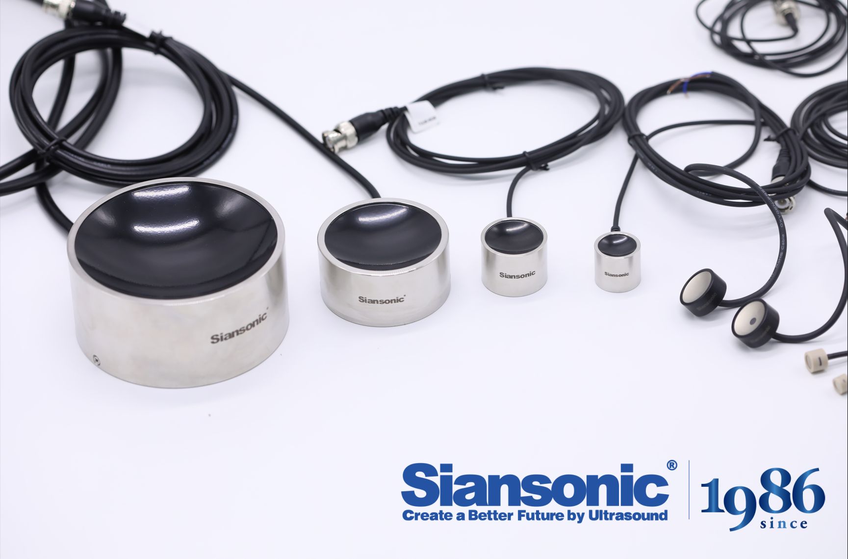
A flat transducer sends acoustic energy forward, but the beam spreads as it travels, losing intensity before it reaches deeper tissue. By curving the active surface or arranging elements along a calculated contour, the focal zone can be pulled inward and tightened, so more of the pulse arrives where it is actually needed. This is not a one-size-fits-all curve; the radius and profile shift depending on tissue depth, expected attenuation, and the size of the region being treated.
Custom geometry also helps correct for the way ultrasound bends at tissue boundaries. Fat, muscle, and bone each have different sound speeds, and a simple spherical bowl will often smear the focus when those layers are uneven. A non-spherical or phased surface can pre-distort the wavefront in the opposite direction, so by the time the beam passes through those irregular layers, it converges cleanly on the target instead of scattering into surrounding healthy tissue.
During design, the surface profile is tuned through repeated simulation and bench testing rather than guessed from a standard template. Each element's tilt, phase delay, and contour are adjusted until the focal spot stays stable across the intended treatment window. That level of geometric control means less energy is wasted, treatment times stay shorter, and the margin between the target and nearby sensitive structures remains safer.
Acoustic Lenses and Phased Arrays: A Smarter Pairing
Acoustic lenses bend sound much like optical lenses bend light, shaping wavefronts without any moving parts or power draw. They excel at creating a fixed focal zone or spreading energy evenly across a broad area, but they lack the ability to adapt in real time. Phased arrays, on the other hand, steer and focus beams electronically by delaying signals across individual elements, giving engineers precise control over direction and depth. Pairing the two seems counterintuitive at first—why combine a passive, fixed device with an active, reconfigurable one? Yet that contrast is exactly what makes the combination work so well.
The lens can act as a coarse, wide-aperture pre-focusing stage, reducing the angular range the phased array needs to cover. This lets the array operate with fewer elements or lower time-delay resolution while still achieving tight beam control. In medical ultrasound, for example, a fixed acoustic lens can set the general depth of field, and the array fine-tunes the focus for different tissue layers. In underwater sonar, a lens can widen the transmit beam for broad search coverage, while the array handles narrow tracking beams on demand.
Another advantage is robustness. Since the lens handles part of the beam shaping passively, the phased array does not need to run at full electronic steering capacity all the time. That lowers power consumption, reduces heat buildup, and can simplify the backend processing. If the electronic control fails partially, the lens still provides a usable acoustic path, which is valuable in harsh environments. The pairing also opens up design flexibility: engineers can swap different lens profiles to change the baseline acoustic behavior without redesigning the entire array.
Reducing Off-Target Heating Without Sacrificing Depth
Achieving deep, uniform thermal effects in tissue without collateral damage is a persistent challenge. The key lies in controlling energy delivery so that the desired target volume reaches therapeutic temperature while surrounding structures remain safely below injury thresholds. This requires precise modulation of pulse duration, power, and cooling intervals, allowing heat to build gradually at depth rather than spiking near the surface.
A practical approach involves pre-cooling the skin and applying intermittent energy bursts. By cycling between active heating and passive cooling, the superficial layers shed excess heat while deeper tissue accumulates it. This temporal staggering exploits differences in thermal relaxation times, letting you push more total energy into the treatment area without raising surface temperature excessively.
Another overlooked factor is the spatial distribution of the energy source. Shifting from a single focal point to a diffuse or multi-lobed pattern spreads surface exposure, reducing peak intensity at any one spot. Combined with real-time temperature feedback, this lets you maintain aggressive depth while automatically throttling back if surface sensors detect an unsafe rise. The result is a treatment that feels less painful, heals faster, and still delivers the deep remodeling effect patients expect.
Bench Tests That Mimic Real Tissue Challenges
Standard bench tests often fall short because they treat tissue as a uniform, passive material. Real surgical sites, however, are anything but uniform—fat, fascia, and muscle each resist needles, sutures, or staplers in distinct ways, and those responses shift with hydration, temperature, and even patient age. To truly gauge how an instrument will perform in the operating room, you need test setups that replicate these layered, living challenges rather than relying on a single slab of silicone.
One effective approach is to build composite tissue phantoms that combine gelatins of varying stiffness, embedded fibrous meshes, and fluid-filled cavities. For example, testing a laparoscopic grasper against a phantom that includes a slippery, low-friction surface followed by a tough, collagen-dense layer exposes weaknesses that a simple pull test would miss—like jaw slipping under oblique loads or tip deflection when encountering sudden resistance. Another common trick is to apply cyclic loading while the phantom is kept warm and moist, mimicking the repetitive motion of a long procedure and the resulting tissue fatigue.
The value of such tests isn't just about passing a spec sheet. When a device can repeatedly withstand forces that mimic a live human body, surgeons gain confidence that it won't tear through delicate structures or lose grip at a critical moment. These bench tests, when designed thoughtfully, become a bridge between abstract material data and the messy, unpredictable reality of surgery.
Where Precision Ultrasound Therapy Goes Next
The next frontier for precision ultrasound therapy isn't just sharper imaging—it's smarter energy delivery. Clinicians are moving beyond static focal points toward adaptive systems that read tissue response in real time, adjusting frequency, pressure, and pulse duration mid-treatment. This closed-loop approach could turn a once-blunt instrument into a scalpel guided by live feedback, reducing collateral heating and opening doors to targets once considered too risky.
Another shift is happening in transducer design. Flexible, multi-element arrays are replacing rigid single-crystal probes, allowing beam steering across irregular anatomical contours without repositioning the patient. Combined with miniaturized electronics, these arrays could bring ultrasound therapy to outpatient settings and even wearable devices, where chronic pain or deep-tissue lesions are managed continuously rather than in isolated sessions.
Finally, the integration of machine learning models trained on acoustic signatures is redefining what "precision" means. Instead of relying solely on operator skill, systems are learning to distinguish between healthy and pathological tissue from backscatter patterns, then automatically sculpting the treatment envelope. The result is a therapy that no longer just targets coordinates, but understands the tissue it touches.
FAQ
A custom unit is shaped around the specific acoustic pathway and target volume rather than forcing clinicians to adapt to a generic focal geometry. It often includes patient- or application-specific curvature, element spacing, and frequency tuning, which lets the beam converge more tightly on the intended tissue while steering energy away from sensitive structures.
By matching the transducer's aperture and phasing to the actual anatomy, the design reduces phase aberrations caused by bone or fat layers. The result is a sharper focal spot with less drift, so the delivered energy stays within the planned treatment margin even when the target sits near critical nerves or vessels.
Simulations allow engineers to model how ultrasound waves traverse different tissue types before any physical device is built. This iterative virtual testing refines element layout and driving signals, which helps avoid hot spots and ensures the focal zone aligns with the clinical target under realistic conditions.
Neurological disorders such as essential tremor and Parkinson's disease see immediate gains because the target volumes are small and surrounded by eloquent brain tissue. Applications like prostate ablation, uterine fibroid treatment, and even blood-brain barrier opening also stand to gain from reduced energy spillover.
Yes, more elements and tailored electronics introduce additional points of failure and require stricter quality control. However, modern fabrication methods and embedded self-test routines have narrowed this reliability gap, making custom designs viable for repeated clinical use without excessive maintenance burdens.
Skull thickness, tissue interfaces, and target depth are not uniform across individuals. Custom designs incorporate imaging data to adjust the transducer's focal length, element distribution, or even the coupling medium, ensuring the beam reaches the planned coordinates without being distorted by irregular bone geometry.
Piezoelectric composites, 3D-printed backing structures, and laser-defined electrode patterns now allow manufacturers to produce complex transducer shapes at lower cost and with better repeatability. These advances also support broader bandwidths, which improves both imaging feedback and therapeutic delivery in the same device.
In principle, yes. When the focal volume is more confined and side lobes are suppressed, less energy reaches surrounding tissue. This lowers the risk of thermal injury or cavitation in off-target regions, though the clinical outcome still depends on accurate targeting and real-time monitoring.
Conclusion
The shift away from stock transducers marks a turning point in focused ultrasound therapy. Rather than relying on generic bowl-shaped elements that scatter energy unpredictably, the custom design reshapes the transducer surface so that each point on the array contributes to a tighter focal zone. This is not just a tweak in curvature; paired acoustic lenses and phased-array controls allow the beam to bend and refocus in real time, compensating for skull aberrations or tissue heterogeneity. By tailoring the geometry to the specific target depth, the system keeps the focal spot sharp without pushing excessive energy into surrounding tissue.
What sets this approach apart is that the precision is validated in bench tests designed to mimic real tissue challenges—heterogeneous phantoms, bone layers, and moving targets. These tests show a measurable drop in off-target heating while maintaining penetration to deep-seated tumors. The smart pairing of lenses and arrays also opens the door to treating regions previously considered too risky, such as close to nerves or blood vessels. Looking ahead, this custom transducer concept could evolve into patient-specific designs generated from preoperative imaging, making focused ultrasound therapy more like a surgical scalpel than a blunt instrument.
Contact Us
Contact Person: Ivy Zhang
Email: [email protected]
Tel/WhatsApp: +86 1081502288
Website: https://www.siansonic.com/